The appearance of Geuda sapphire is a translucent gemstone because of the tiny inclusions widely spreading throughout the stone. These inclusions appear as silky or tiny particles. The stone can be heated to improve transparency and to yield blue color (Schmetzer and Kiefert, 1990; Jaliya et al., 2020). Heat treatment usually affects milky and silky appearances as well as color modification, and even the internal appearance (including inclusions) of corundum.
Several researchers have previously described the technical heat treatment and cause of color in sapphire (e.g., Nassau 1981, Koivula 1987; Fritsch and Rossman, 1987; Schmetzer and Kiefert, 1990; Scarratt, 1999; Jaliya et al., 2020). Moreover, UV-Vis-NIR spectrophotometer is probably useful in the identification of sapphire, and it may also provide information on the geographic origin (Scarratt, 1999). In addition, the absorption spectra have been investigated for cause of blue color in sapphire; they display main broad absorption bands centred at around 570 nm (o-ray) and 730 nm (e-ray), responsible for Fe2+-Ti4+intervalence charge transfer (IVCT) mechanism (Lehmann and Harder, 1970; Häger, 2001).
For the absorption spectrum, optical properties of particle inclusions, commonly found in Sri Lanka sapphire, have been investigated. Mcneil and French (2000) studied various concentrations of TiO2 particles on semi-transparent resin films. The absorption spectra showed the scattering of TiO2 particles by the absorption edge below 400 nm. Moreover, Popov (2008) suggested that the TiO2 nanoparticles can be used as the UV protectors to skin because the nanosized TiO2 spheres, ranged from35-200 nm in diameter, can scatter the UV radiation (310 and 400 nm). According to the tiny inclusion in Geuda sapphire, the absorption edge might be found because of the scattering. Therefore, the milky Geuda sapphires under this experiment were not only investigated regarding the color created by heat treatment but also the influence of tiny inclusions in absorption spectrum.
Materials And Methods
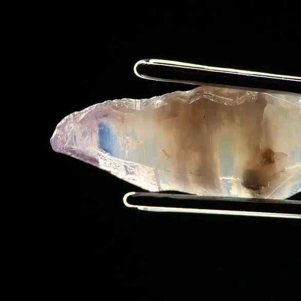
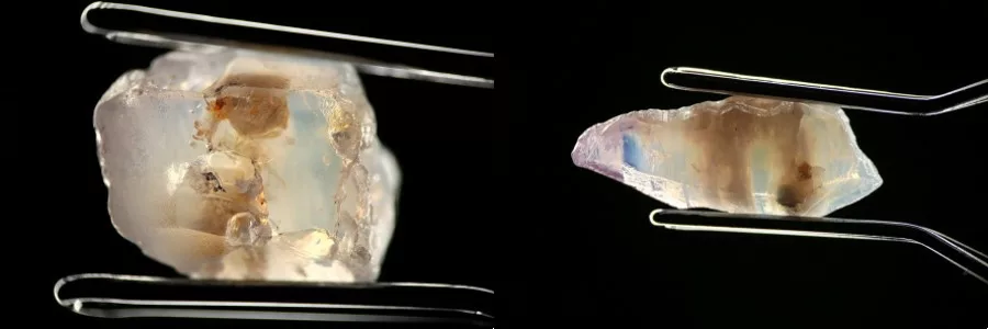
There were 10 rough samples of unheated Geuda sapphire from Sri Lanka in this study The color of the samples was light-blue color with milky appearance. After sample preparation, cutting, and polishing both sides to be slab (without crystal orientation), they were showed clearly silk and cloud inclusions in all samples (Fig. 1). Electron Probe Micro-Analyzer (EPMA) JEOL model JXA-8100 was engaged for chemical analyses of major and trace element compositions of host sapphire samples. UV-Vis-NIR absorption spectra were measured by the VARIAN Cary 500 UV-VIS-NIR spectrophotometer with the wavelength range 175-3300 nm. Samples were heated by Linn electrical furnace (model HT 1800 Plus VAC Bottom Loader) at a temperature of 1650°C with 5 hours soaking time. The atmospheric condition in the chamber was controlled by pure N2 gas feeding throughout the heating and cooling processes.
Results and Discussion
Physical properties:
The samples were typically semi-transparent to transparent appearance. Their RIs fall within 1.760–1.771 range, SG values are 3.90-4.00 with averaged birefringence of 0.008– 0.010. All samples are inert fluorescence in both long-wave and short-wave UV radiation.
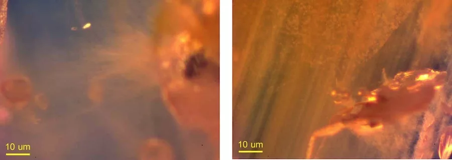
Inclusions of Geuda sapphire samples from Sri Lanka show a feature of milky appearance. In this study, the samples can be divided into two groups, based on inclusion appearance, i.e., silk and cloud. Silk inclusions appear intersecting of silk along the crystal structure. Cloud inclusions include minute particles dispersing throughout the sample. Both types of inclusions can be observed clearly under the gemological microscope which their sizes are less than 1 µm and disperse at a distance less than 1 µm (Fig. 2).
Absorption Spectra:
The absorption spectra of sapphire normally show the absorbion bands of Fe3+ at 377, 388, and 450nm (Emmett, et al. 1993). Whereas the milky Geuda yields the absorption edge pattern in a range of 300 – 350nm. This absorption edge is probably caused by the amount and size of the inclusions (e.g., silk and cloud) in Geuda sapphire as previously reported at wavelength lower than 400 nm (Mcneil and French, 2000; Egerton, 2014).
After the heat treatment at 1650°C under a reducing atmosphere, Geuda sapphires turn whitish color to blue color. The absorption spectra show increasing band at around 560 nm, corresponding to the intervalence charge transfer in the form of Fe2+ – Ti4+ (IVCT). However, the absorption edge is vanished away due to the dissolution of minute particles during heat treatment (Fig. 3).