Detection of low-temperature heat treatment (below 1200°C) in ruby and sapphire can present challenges to gemologists, as alterations to the material are often subtle. In this study, the authors heated Burmese ruby samples from Mogok, Myanmar, at temperatures ranging from 600° to 1500°C. The samples were documented using macrophotography, photomicrography of inclusions, ultraviolet fluorescence imaging, and spectroscopic analysis (Raman, ultraviolet/visible/near-infrared, and infrared) to record any changes, with a focus on features that could help detect heat treatment. A wide variety of solid inclusions, including calcite (Figure 1), mica, spinel, and zircon, were found in Mogok ruby. Many of those were found to be sensitive to heat treatment with regard to morphology and phase transition; their reactions varied depending on a number of factors such as size, distance from the surface, and species. Microscopic examination provided useful visual indications of heat treatment, even at lower temperatures (Figure 2). Raman analysis of calcite and spinel inclusions (Figure 3) also proved valuable in providing complementary evidence of low-temperature heat treatment. Spinel inclusions in particular showed a widening of peak in the range of 405–420 cm–1, which is known to occur when spinels are heated (Saeseaw, 2009). Fluorescence images were also recorded before and after treatment (Figure 4). The combination of these factors can assist gemologists in detecting low temperature heat treatment.
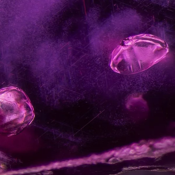
Figure 1: A pair of calcite crystals stands out above a cloud of undissolved rutile silk needles. Note the twinning planes in both crystals, a common feature for calcite. Photomicrograph by E. Billie Hughes; field of view 2 mm.
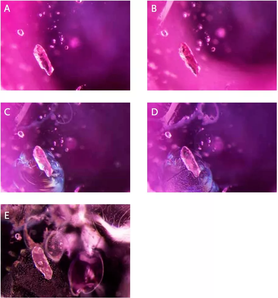
Figure 2. A: (Unheated) An elongated mica crystal surrounded by several smaller crystals in sample 8. B: (600°C) There is little noticeable change except at the top left, where a small fissure has formed. C: (900°C) The scene shows obvious signs of heat alteration. In addition to the fissures seen earlier, glassy discoids appear around crystals in the background, and a shiny fissure is conspicuous around the biotite crystal in the foreground. D: (1100°C) Some fissures have begun to heal; the glassy fissure around the biotite crystal has developed elongated channels, and the partially healed edges of the glassy discoid at the top of the frame have started to neck down and become wider. E: (1500°C) The scene has changed dramatically; glassy discoids and partially healed fissures with a melted appearance are visible across the field of view, and the biotite crystal has developed an immobile gas bubble in its center. Images A and B were captured with darkfield illumination; diffused fiber-optic illumination has been added in C–E to reveal the fissures more clearly. Photomicrographs by E. Billie Hughes; field of view 1.5mm.
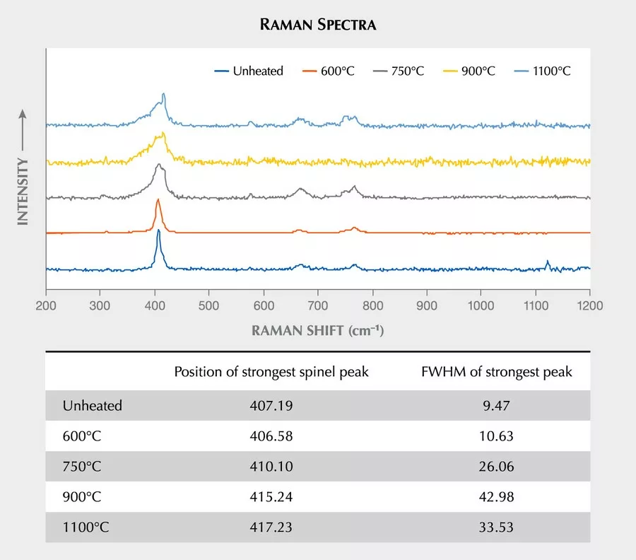
Figure 3. Raman spectra of a spinel inclusion in one of the heated ruby samples. After the first round of heating to 600°C, the spectrum remained consistent, but notable changes occurred after heating to 750°C. The peak between 405 and 420 cm–1 widened and shifted to a slightly higher wavenumber, a trend that continued with heating to higher temperatures. Spectra are offset vertically for clarity.
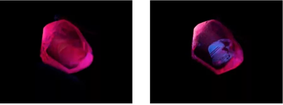
Figure 4. A: Sample 46 displays a strong red short-wave fluorescence in its unheated state. Like the other samples tested, it did not display any change in appearance in short-wave UV illumination until after the last round of heating. B: After heating to 1500°C, the ruby displays a clear, zoned chalky appearance in short-wave UV. Photos by E. Billie Hughes.