Influence of Irradiation on Colour Modification and Colour Stability of Rubies: A Preliminary Study

By Chanenkant Jakkawanvibul, Chotika Kittikunlayaworakun, Kanwalee Pangza, Nongnuch Jangsawang, Thanapong Lhuaamporn, Thanong Leelawatanasuk, Visut Pisutha-Arnond, Waratchanok Suwanmanee, Wilawan Atichat

Keywords: Ruby, Treatment, Gamma radiation, Electron radiation, Colour stability test

Figure 1. Gem-quality rubies from three different and important sources (left: Madagascar, 1.02 ct; middle: Myanmar, 1.52 ct; right: Mozambique, 1.18 ct. Photo by W. Suwanmanee.

Introduction

Ruby is the chromium-bearing red colour-variety of corundum Al2 O3 . Since historic times, ruby is highly popular in the gem and jewellery market because of its highly saturated red colour, high hardness and brilliancy (Figure 1). In recent years, the demand for rubies has continuously increased while production of fine quality stones is getting less. Therefore, some suppliers are preferring to improve rubies of lower quality by various heat treatments to enhance mainly their colour and clarity. In addition to heating, there is also irradiation treatment on corundum. Pough & Rogers (1947) reported that X-rays can produce yellow colour hues in all varieties of corundum but that the extent of colour shift after irradiation is also dependent on the original colour saturation of the stones.

In case of rubies, there are some methods to remove their purplish tint and turn them to a brighter red colour which is achieved by the addition of a yellow hue such as low temperature heat treatment and Be heat treatment. For irradiation method, it is known that the yellow coloration after irradiation of corundum is related to defect structures (colour centres) and that they may be unstable to extensive exposure to daylight and/or thermal energy (Pisutha-Arnond et al., 2004). This is specifically the case when the colouration after being irradiated is unstable and fades back into the original colour.

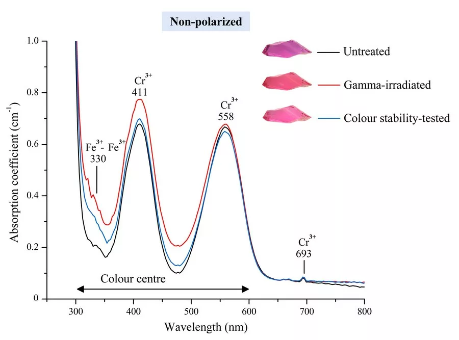
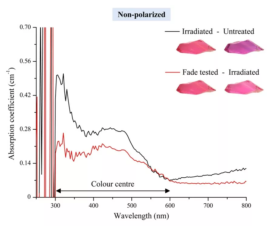
Therefore, the authors have investigated a selection of irradiated rubies to better understand the effect of this treatment and to develop potential detection methods for gem testing laboratories. In the present study, selected rubies of purplish red colour from Myanmar, Mozambique, and Madagascar were investigated. Gamma and electron beam irradiation experiments on our ruby samples were carried out in a specialized facility in Thailand . All rubies were subjected to a colour stability test after being irradiated. The colour modification of the ruby samples was investigated by UV-Vis absorption spectra and colour photographs to compare their colour before and after irradiation and after the colour stability test.

Materials and Methods

Six untreated rubies were selected for the present study, consisting of rough and faceted samples. They are from three sources: Myanmar, Mozambique, and Madagascar. All samples were provided by the Gem Testing Laboratory of the Gem and Jewelry Institute of Thailand (Public Organization).

Photographs of the samples were taken in a standard light box with 5500K Fluorescence light bulbs. Colour stability test of the rubies after irradiation was done using fiber optic lamp (halogen) at 15V and 150 W. Exposure time for testing was 6 hours.

Gamma and electron (e-beam) radiations were applied for ruby treatment at a dosage of 1,000 kGy. Gamma radiation was performed using six columns of cobalt-60 (60Co) at an energy of 1.17 and 1.33 MeV. For electron beam irradiation we used a high-energy electron accelerator operated at 20 MeV with a power of 10 MW. The facility for both irradiations is located at The Thailand Institute of Nuclear Technology (Public Organization), Nakhon Nayok province. The UV-Vis absorption spectra for rubies before and after treatment were analyzed using a PerkinElmer LAMBDA 1050 spectrophotometer. The result of absorption intensity was reported in absorption coefficient (cm-1) units.

Results of irradiation treatment

Colour modification by irradiation and colour stability test
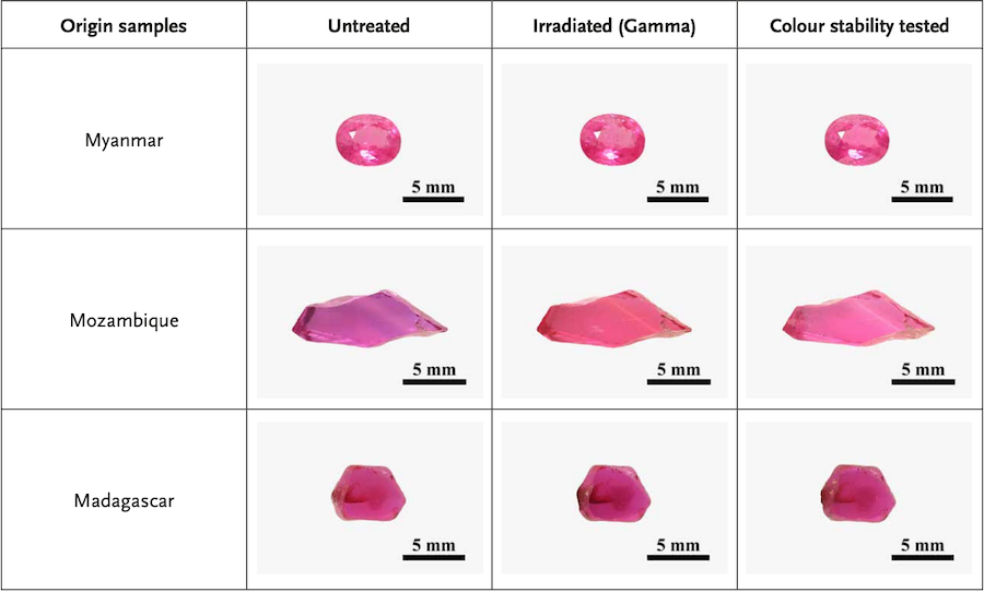
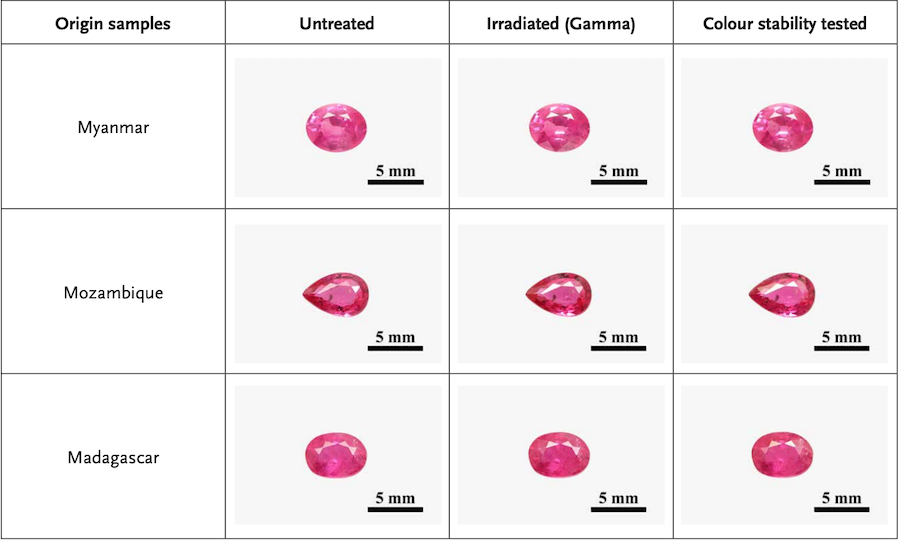
Gamma radiation: Figure 2 shows the colour of rubies before and after irradiation, and also after colour stability testing. Gamma radiation produced a slight yellow tint in Mozambique ruby. This resulted in an orangey red colour of the sample. In addition, the blue colour banding disappeared. The colour of our Madagascar sample became slightly darker red after irradiation, whereas the ruby from Myanmar remained unchanged by the gamma-irradiation. After colour stability testing, the yellowish tint in the irradiated Mozambique ruby was removed and the sample turned to pinkish red, a distinctly brighter colour than before irradiation. In contrast to this, the ruby samples from Myanmar and Madagascar remained unchanged after colour stability testing.

Figure 2: Colour modification of rubies after irradiation with gamma radiation and after colour stability test. Photos by W. Suwanmanee.

Electron radiation (e-beam): After electron beam irradiation, the additional ruby samples from Mozambique and Madagascar turned to a very slightly brighter red, whereas the ruby sample from Myanmar remained unchanged. All samples remained unchanged after being tested for colour stability (see in Figure 3.).

Figure 3: Colour modification of rubies after irradiation with an electron beam and after colour stability test. Photos by W. Suwanmanee.

UV-Vis spectroscopy
The absorption spectrum of ruby is commonly dominated by two broad Cr3+-related bands centered at about 411 and 558 nm and an additional series of small Cr3+ peaks at 693 nm (Schmetzer & Schwarz, 2004; Promwongnan & Sutthirat, 2019). An additional band at around 330 nm is due to the presence of Fe3+ – Fe3+ pairs (Promwongnan & Sutthirat, 2019). The absorption spectra of the ruby sample from Mozambique (Figure 4) reveals the increase of the absorptivity in the range of 300 – 600 nm after gamma-irradiation and a slight decrease after the stability testing. By subtracting the initial untreated UV-Vis absorption spectrum from the irradiated spectrum (Figure 5, black line) this increase in absorption after irradiation becomes even more evident (two broad bands between 300 – 600 nm) likely as a result of an activated colour centre (Pisutha-Arnond et al., 2004). However, these irradiation-induced bands were slightly reduced after being tested for color stability (Figure 5, red line). 

Figure 4: Comparison of the UV-Vis absorption spectra of the Mozambique ruby sample before treatment (black line), after being irradiated with gamma radiation (red line), and after colour stability testing (blue line).
Figure 5: Resulting UV-Vis spectra after subtracting the initial untreated spectrum from the gamma-irradiated (black line), and the spectrum after colour stability test from the gammairradiated spectrum (red line).

Concluding remarks

The experimental results show that both gamma- and electron beam radiation may produce (a slight) yellowish colour hue in purplish red ruby, particularly in those from East-Africa (e.g., Mozambique and Madagascar). Our experiments have shown that gamma radiation can produce more pronounced yellowish colour hues than electron beam radiation. The slight yellow colour shift after irradiation results in a slight increase of absorption between 300 – 600 nm, caused by an activated defect structure (colour centres). By applying a colour stability test we were able to remove the yellowish hue partially in the irradiated ruby, specifically in the Mozambique ruby. But the colour in this sample did not fully revert to the original purplish red colour. As shown, the absorption spectrum may change slightly by this treatment. However, this slight spectral change can so far not be able to use safely as an indication that the ruby has been treated by irradiation. More experiments must be carried out in the near future to better understand irradiation treatments of rubies and its colour stability.

References:

  • Pisutha-Arnond, V., Hager, T., Wathanakul, P., and Atichart, W. 2004. Yellow and Brown Colouration in Beryllium-Treated Sapphires, Journal of Gemmology, 29 (2), 77-103.
  • Pough, F.H. and Rogers, T.H. 1947. Experiments in x-ray irradiation of gemstones. American Mineralogist, 32 (1-2), 31–43.
  • Promwongnan, S. and Sutthirat, C. 2019. An Update on Mineral Inclusions and Their Composition in Ruby from the Bo Rai Gem Field in Trat Province, Eastern Thailand, Journal of Gemmology, 36 (7), 634-645.
  • Schmetzer, K. and Schwarz, D. 2004. The Causes of Colour in Untreated, Heat Treated and Diffusion Treated Orange and Pinkish-Orange Sapphires – A Review, Journal of Gemmology, 29 (3), 149-182.

Acknowledgement
This work was funded by the Gem and Jewelry Institute of Thailand (Public Organization) for fiscal budget in 2022 and was supported by Thailand Institute of Nuclear Technology (Public Organization) for irradiation treatment.